We have been exploring the fascinating phenomenon whereby amoeboid cells of the immune system can crawl upstream against the direction of flow through LFA-1/ICAM-1 interactions. In these experiments, Sarah Kim showed that T-cells can remember the direction of upstream migration after the flow is turned off, if they are on surfaces that present both VCAM-1/ICAM-1 surface even after the flow is turned off. (SHJ Kim and DA Hammer. Integrative Biology, 2019) Alex Buffone in our lab has also recently shown that hematopoietic stem cells can crawl upstream, and neutrophils can do so also if Mac-1 is blocked. The neutrophil work is published here: https://www.sciencedirect.com/science/article/pii/S0006349519307908.
We recreated the rolling/firm/adhesion/upstream migration cascade in a purified molecular system by printing a chemokine (SDF-1alhpa, ICAM-1 and E-selectin on a surface). T-lymphocytes roll to a stop, are activated, and migrate upstream. It’s fascinating to watch! Video taken by Nick Anderson.
PROTOCELL ENGINEERING
We constructed motile, adhesive protocells by encapsulating catalase in polymersomes that were adherent to a surface via low density biotin-avidin interactions. When hydrogen peroxide is added to the solution, it crosses the polymersome membrane, and is converted by catalase and generates a force which propels the protocells. The motion is diffusive (because there is no gradient of hydrogen peroxide). Work done by Woo-Sik Jang, in collaboration with the Lee lab, published in Small http://doi.org/10.1002/small.2018201715.
ADHESIVE DYNAMICS
The Hammer lab has developed adhesive dynamics, a computational tool for simulating adhesion in many biological circumstances, including leukocyte homing and viral binding. A good review can be found at this link (https://doi.org/10.1115/1.4026402). In the above simulation, we combined adhesive dynamics with an intracellular signaling pathway to simulate the progressive rolling and stopping of a T-lymphocyte on a ligand coated surface. LFA-1 integrins are converted to a high affinity state (shown in blue on the cell) and ultimately, the cell comes to a stop when sufficiently many LFA-1 molecules are activated. The inset shows the reduction in rolling velocity as the cell is activated. Simulation performed by Kelly Caputo; original development of integrated signaling adhesive dynamics done by Mike Beste and Ellen Krasik.
CONTROLLING VESICLE ASSEMBLY AND ENCAPSULATION
In collaboration with Daeyeon Lee (CBE, Penn), we can assemble micron-scale soft matter materials of defined size and composition, to include active biological molecules, control switches, membraneless organelles and active enzymes.
MEMBRANELESS ORGANELLES FROM RECOMBINANT PROTEINS
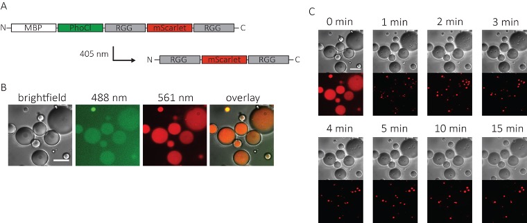
We constructed light-activated membraneless organelles using the recombinant protein PhoCl attached to RGG dimers, which Ben Schuster had previously engineered to coacervate. Ellen Reed inserted PhoCl between the solubilizing maltose binding protein and RGG-dimer, with an integrated mScarlet as a reporter tag. After illumination with light at 405 nm, PhoCl is cleaved and RGG-dimers form membraneless organelles! Work done in collaboration with Matt Good. https://pubs.acs.org/doi/abs/10.1021/acssynbio.9b00503
VESICLES AND OTHER STRUCTURES FROM RECOMBINANT PROTEINS
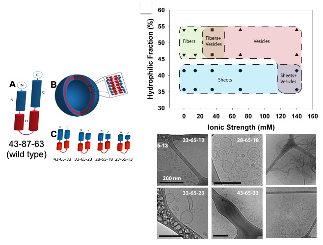
We have constructed membranes (and other soft matter structures such as micelles and sheets) from a recombinant protein called oleosin. Kevin Vargo showed that a small family of engineered oleosin variants can be assembled to different soft matter structures, as a function of the size of the head groups and the ionic strength of the solvent. https://www.pnas.org/content/early/2012/06/25/1205426109.